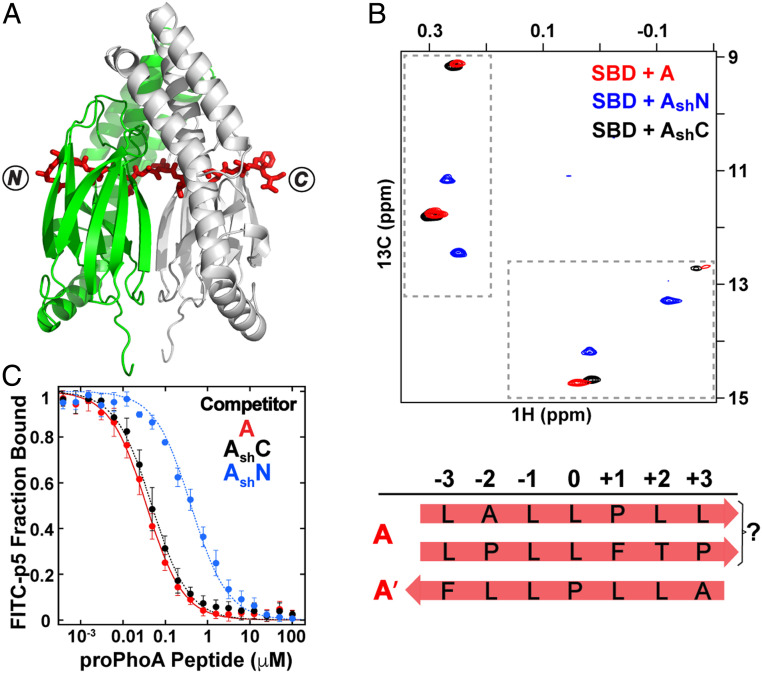
Fig. 6.
The peptide containing proPhoA site A spans an SBD dimer in crystals and in solution binds 1:1 to the SBD, populating more than one binding mode. (A) Crystal structure of the DnaK SBD in complex with peptide A. Two SBDs (green and gray) are aligned at the twofold axis, and peptide A (red sticks) extends through both SBDs with two sequences in the binding clefts, one in an N to C and one in a C to N backbone direction. (B) The Ile401 and Ile438 region of 1H-13C-HMQC spectra of ILV-13CH3–labeled DnaK SBD in the presence of unlabeled peptides A (red), AshN (blue), and AshC (black). The binding modes were assigned as 1) N to C with a leucine in the central pocket and 2) C to N with P(−10) in the central pocket. (C) Competition binding assays for peptides A, AshN, and AshC to the SBD bound to a fluorescein isothiocyanate (FITC)-labeled peptide (SI Appendix, SI Materials and Methods and Fig. S10).

