Fig. 11.
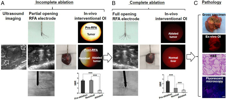
Preclinical longitudinal validation of the technical feasibility of the interventional OI. (A) Ultrasound imaging detected the tumor (between open arrows). An incompletely ablated tumor was purposely created, and the interventional OI with quantifying SBR analysis was used to instantly detect the residual tumor, which demonstrated high fluorescence signals (yellow). (B) Based on findings of the interventional OI, the ablation was repeated with a larger ablation zone and increased energy to specifically destruct the residual tumor. Thus, the intraprocedural real-time OI guidance ensured the final complete tumor ablation, which demonstrated a low fluorescence signal in the ablated tumor. (C) Ex vivo OI and subsequent pathology with different examinations finally confirmed complete tumor eradication (necrosis). [Scale bar (H&E), 20 μm; scale bar (fluorescent microscope), 50 μm.] A paired Student’s t test was used for statistical analysis (***P < 0.001).

