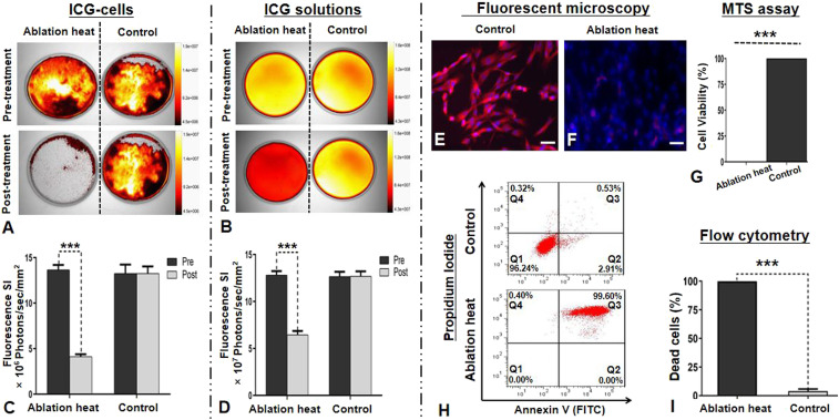
Fig. 4.
In vitro OI for the evaluation of ablation heat effect on ICG-treated VX2 tumor cells (ICG cells) and ICG solutions. (A and B) The optical/X-ray images demonstrated clearly the decreased fluorescence signals (yellow-orange) of both ICG cells and ICG solutions after the lethal ablation heat treatment at 80 °C, while no signal changes were noted in the non–ablation-heat control groups. (C and D) Quantitative analysis further confirmed a significant decrease in the fluorescence SIs of the ablation-heated ICG cells and ICG solutions, with no significant SI changes in the controlled ICG cells and ICG solutions. (E and F) Fluorescent microscopy images further displayed that fluorescence signals of ICG cells were clearly decreased with smudged nuclei after the ablation heat treatment, compared with the non–ablation-heated control cells. (G) The MTS (3-(4,5-dimethylthiazol-2-yl)-5-(3-carboxymethoxyphenyl)-2-(4-sulfophenyl)-2H-tetrazolium) assay showed no viable cells after the ablation heat treatment. (H and I) Flow cytometry further confirmed almost 100% nonviable cells (red in Q3 zone) after the ablation heat treatment, compared with the viable cells (red in Q1 zone) of the control group without ablation heat treatment. (Scale bars, 50 μm.) A paired Student’s t test was used for statistical analysis (***P < 0.001).

