Fig. 5.
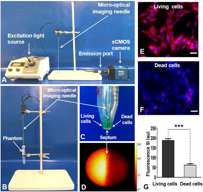
(A) The interventional OI system. Because of the designed needle type, the micro-OI needle could be inserted to the tumor via a percutaneous access to achieve an “inside-output” real-time intratumoral imaging. (B and C) In vitro experimental setup for evaluating the interventional OI system. A cell phantom was created, consisting of a plastic tube that had two separate chambers in the inferior portion (C). The living (viable) tumor cells were centrifuged in one chamber, with nonviable tumor cells (treated by 80 °C heat) in the opposite chamber. The micro-OI needle was vertically positioned in the phantom, just above the two cell-containing chambers. (D) Real-time interventional optical image, demonstrating a high ICG signal (yellow) for the living cell group compared with the dead cell group, which was further confirmed by fluorescent microscopy (E and F) and fluorescent SI quantification (G). (Scale bars, 50 μm.) A paired Student’s t test was used for statistical analysis (***P < 0.001).

