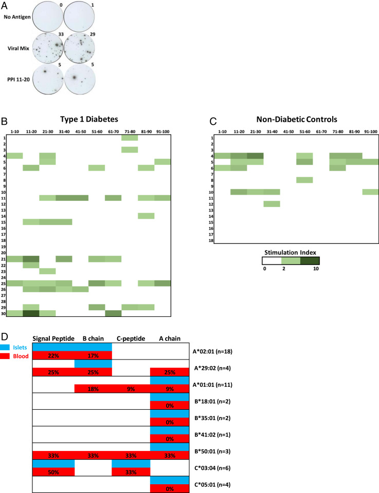
Fig. 6.
Peripheral blood immune responses to preproinsulin (PPI) peptide pools. In each case, cryopreserved PBMCs were cultured in the presence or absence of peptides for 48 h, washed, and then cells transferred to an IFN-γ monoclonal antibody–coated plate for overnight culture followed by development and enumeration of ELISPOTs. (A) Representative IFN-γ ELISPOT images from PBMCs stimulated with no antigen, a viral mix of peptides, and preproinsulin peptide pools containing amino acids 11 to 20. Heat maps showing the response of PPI pools in (B) new-onset T1D patients (n = 30) and (C) nondiabetic subjects (n = 18) as a stimulation index (SI) (No. ELISPOTs in a PPI pool / No. ELISPOTs no antigen). (D) Comparison of islet-derived TCR clonotypes responding to PPI epitopes from T1D organ donors to those from the peripheral blood of new-onset T1D subjects by HLA class I association. Percentages depict the number of new-onset T1D subjects with a given HLA allele that had a SI ≥ 3 for peptide pools within a region of preproinsulin; n = number of new-onset T1D patients with a given HLA allele. White indicates no reactivity found in islets from T1D organ donors or peripheral blood of new-onset subjects.

