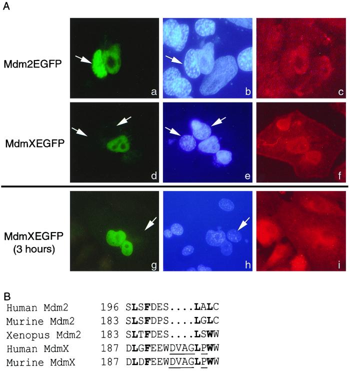
FIG. 3.
MdmX is unable to undergo nuclear export. (A) Fluorescence of Mdm2-EGFP (a) or MdmX-EGFP (d and g) is observed in three heterokaryons. The human and murine nuclei (b, e, and h, white arrows) are visualized by Hoechst dye. Murine nuclei are indicatively stained with a punctate pattern distinct from that of the human nuclei. A β-actin antibody followed with a Texas red-conjugated secondary antibody allowed confirmation that the nuclei are within heterokaryons (c, f, and i). Mdm2-EGFP serves as a positive control demonstrating that the EGFP fusion does not inhibit nucleocytoplasmic shuttling. The data presented are representative of more than 20 separate heterokaryons. (B) Comparison of the Mdm2 NES with the putative MdmX NES. Boldfaced amino acids represent conserved hydrophobic amino acids; underlined amino acids produce an altered secondary structure within the MdmX NES.

