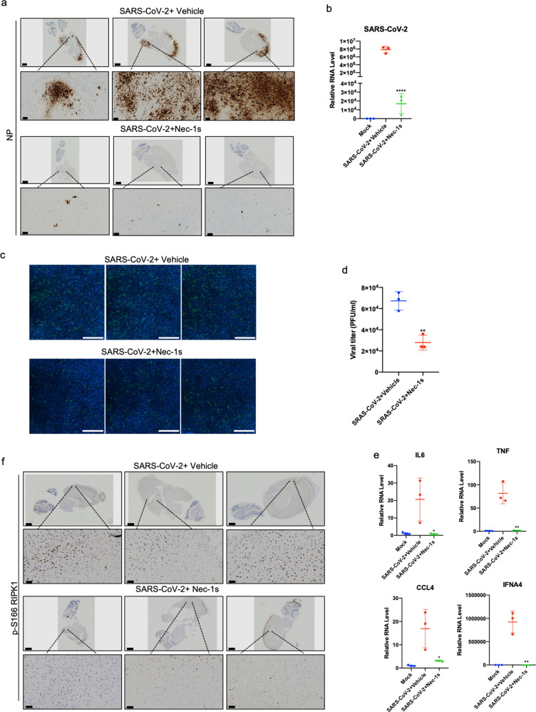
Fig. 6. Nec-1s treatment reduces the viral loads in the brain of AC70 transgenic mice.
a IHC staining with antibody against viral NP on the saggital whole brain sections of the SARS-CoV-2-infected AC70 transgenic mice with or without Nec-1s treatments. Scale bars, 1000 μm. The lower row shows the enlarged images of specified areas above. Scale bars, 100 μm. Viral NP was robustly detected in multiple brain regions, including the olfactory bulb, the deep cortical layers, the connected hippocampal subiculum, the hindbrain/medulla and the cerebellar dentate nucleus. b Freshly isolated mouse brains were extracted with Trizol for isolating RNA. After reverse transcription, the virus RNA was analyzed by RT-qPCR. Paired t-test was used in RT-qPCR analysis (****P < 0.0001). c, d The brain tissues of viral-infected AC70 mice with or without Nec-1s treatment (1 g) were cryogenically ground in 1 mL DMEM medium. After fully grinding and centrifugation, the supernatant was collected and stored at –80 °C. 4 × 10⁴ Vero-E6 cells were seeded in 96-well plates for 23 h. The supernatant (50 μL) was added to the cells and incubated for 24 h. The SARS-CoV-2 infection was detected by immunofluorescence using COVID-19 convalescent sera (c). Scale bars, 1000 μm. Infectious clones are automatically quantified by Cytation 5 (d). Paired t-test was used in viral titer analysis (**P < 0.01). e The brains of mice in the control group (n = 3) and Nec-1s-treated group (n = 3) were extracted with Trizol for total RNA, and the expression of cytokines was analyzed by RT-qPCR. Paired t-test was used in RT-qPCR analysis (*P < 0.05, **P < 0.01). f RIPK1 was activated in the brains of AC70 transgenic mice with SARS-CoV-2 infection. IHC staining of p-S166 RIPK1 in the brains of control or Nec-1s-administrated AC70 transgenic mice infected with SARS-CoV-2 was shown. Scale bars, 1000 μm. The lower row shows the enlarged images of specified areas above. Scale bars, 50 μm.

