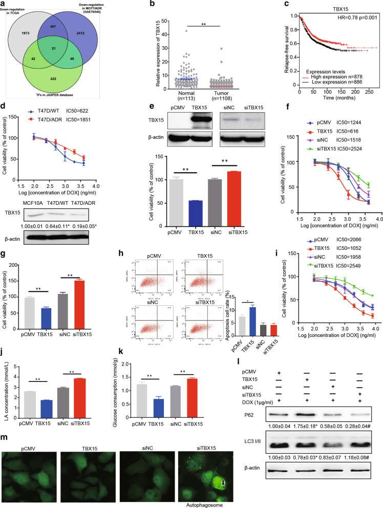
Fig. 1.
TBX15 reduced DOX resistance in breast cancer. A Venn diagram showing the intersection of genes identified in TCGA that are downregulated in breast cancer, genes downregulated in MCF7/ADR cells (GSE76540 database), and from TFs identified in the JASPER database. B Relative gene expression of TBX15 in normal tissue or breast tumors. Gene expression data were obtained from 1108 breast tumors and 113 normal adjacent tissues from TCGA dataset and were presented as the means ± SEM. C Kaplan–Meier plot predicting the Relapse-Free Survival of patients with high or low expression of TBX15. D T47D/ADR cells were developed from T47D/WT cells, and the IC50 values of DOX treatment were tested. TBX15 expression in MCF10A, T47D/WT and T47D/ADR cells was detected in Western blotting assays. β-actin was used as internal reference. E–N Breast cancer cells were transfected with pCMV, pCMV-TBX15, siNC, or siTBX15. E The T47D/ADR cells with different treatments were sent to MTT assays. F Drug sensitivity analysis after DOX treatment in various T47D/ADR groups. G The MCF7/ADR cells with various treatments were sent to MTT assay. H The MCF7/ADR cells with various treatments were sent to flow cytometry to test cell apoptosis rate. I Drug sensitivity analysis after DOX treatment in MCF7/ADR cells. J–K Lactate acid (LA) production and glucose consumption were detected in various T47D/ADR groups. L P62 and LC3 II/I expression in MCF7/ADR cells were detected in Western blotting assays. β-actin was used as internal reference. M The MCF7/ADR cells were transfected with EGFP-LC3 plasmid, and then exhibited various treatments. Fluorescence microscope was used to investigate the autophagy process (400×magnification, Arrows: autophagosomes accumulated with LC3). Data are presented as the means± SEM from three independent experiments. *, **P < 0.05, P < 0.01, respectively. # indicates significant difference compared to siNC group at P < 0.05

