Fig. 6.
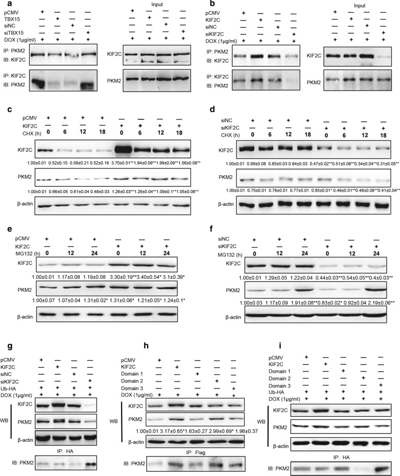
KIF2C directly binds to PKM2 and promotes PKM2 stability in breast cancer cells. A Co-IP analysis of MCF7/ADR cells that were transfected with pCMV, TBX15, siNC or siTBX15 and were treated with 1 µg /ml DOX for 48 h. B Co-IP analysis of MCF7/ADR cells that were transfected with pCMV, KIF2C, siNC or siKIF2C. Cell lysates were co-immunoprecipitated using an anti-KIF2C or anti-PKM2 antibody. C Western blotting analysis of MCF7/ADR cells transfected with pCMV or KIF2C and treated with 20 µg /ml CHX for 0, 6, 12 and 18 h. D Western blotting analysis of MCF7/ADR cells transfected with siNC or siKIF2C and were treated with CHX. The expression levels of KIF2C and PKM2 were analyzed. E Western blotting analysis of MCF7/ADR cells transfected with a pCMV or KIF2C and treated with 1 µM MG132 for 0, 12 and 24 h. F Western blotting analysis of MCF7/ADR cells transfected with siNC or siKIF2C and treated with MG132. The expression of KIF2C and PKM2 were analyzed. G Western blotting analysis of MCF7/ADR cells transfected with pCMV, KIF2C, siNC or siKIF2C and Ubiquitin-HA plasmid. Cell lysates were co-immunoprecipitated using anti-HA antibody and the expression levels of Ub-PKM2 were analyzed. H Western blotting analysis of MCF7/ADR cells transfected with a pCMV, KIF2C, and KIF2C domains (D1, D2, D3) in pCMV-flag plasmids. Cells were treated with 1 µg /ml DOX for 48 h. Cell lysates were co-immunoprecipitated using anti-flag antibody and the expression levels of PKM2 were analyzed. I Western blotting analysis of MCF7/ADR cells that were transfected as (H), and Ubiquitin-HA plasmid. Cell lysates were co-immunoprecipitated using an anti-HA antibody and the expression levels of Ub-PKM2 were analyzed. All the data above were presented as the means ± SEM from three independent experiments and were normalized to the percentage of corresponding control groups. *, ** P < 0.05, P < 0.01, respectively

