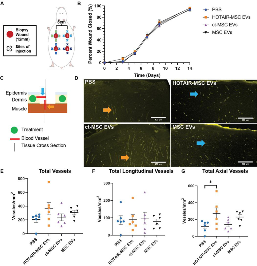
Figure 4. HOTAIR-MSC EVs do not improve healing of excisional wounds in healthy Sprague-Dawley rats but do induce local angiogenic effects.
(A) Schematic of 12 mm excisional wound healing model in Sprague-Dawley rats. (B) Wound closure as assessed using digital planimetry for wounds treated with HOTAIR-MSC EVs, ct-MSC EVs, native MSC EVs, or PBS. Statistical significance was calculated using two-way ANOVA with Holm-Sidak’s multiple comparison test (n=11). (C) Schematic of day 21 tissue processing indicating blood vessel perspective based on the sectioning of tissue with axial vessels originating from the dermis and vertical, longitudinal vessels originating from the underlying muscle. Treatments were injected within the dermis. (D) CD31+ immunohistochemistry of tissue highlighting newly formed blood vessels in the center of the healed wound. Color-coded arrow heads correspond to (C). (E) Total, (F) longitudinal, and (G) axial vessel were counted and displayed as number of vessels per square millimeter of tissue. Statistical significance was calculated using one-way ANOVA with Holm-Sidak’s multiple comparison test (* p<0.05; n=6).

