Fig. 3.

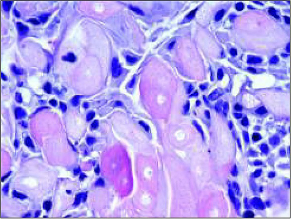
H&E, objective x 63. This high magnification picture shows in detail the large eosinophilic cells as well as the collection of amyloid-like material.
Ryc. 3. Barwienie hematoksylina i eozyna, obiektyw x63. Na tym dużym powiększeniu widoczne są w szczegółach komórki z eozynochłonną cytoplazmą oraz amyloidopodobna substancja.
