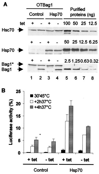
FIG. 2.
Analysis of Bag1 activity on Hsp70 in a regulated cell line. (A) Western blot analysis of Hsc70, Hsp70, and Bag1 expression levels. OTBag1 cells were transiently transfected with a plasmid encoding cytoplasmic luciferase and pCDNA (control) or pCMV70 (Hsp70). At 24 h after transfection, the cells were grown in medium with or without tetracycline (tet) for induction of Bag1 expression. Protein levels in cell lysates (lanes 1 to 4) were compared to levels of purified Hsp70 and Bag1 (lanes 5 to 8). Levels of Hsc70, Hsp70, endogenous Bag1 (Bag1*; also recognized by polyclonal anti-BagΔC antibody), and mouse Bag-1 were 11.5 ± 2.7, 17.3 ± 6.1, 2.0 ± 0.7, and 1.6 ± 0.4 μM, respectively (averages ± standard errors of the means of three independent measurements). Hsp70 levels were corrected for transfection efficiencies that were determined by indirect immunofluorescence. (B) OTBag1 cells were transfected with pCytluc and pCDNA or pCMV70 as described above. After pretreatment with cycloheximide (20 μg/ml), the cells were heated at 45°C for 30 min. During a subsequent recovery period at 37°C, samples were taken at 2 and 4 h after heat shock and assayed for luciferase activity. The data points represent averages of three independent experiments. Error bars indicate the standard errors of the means.

