
FIG. 4.
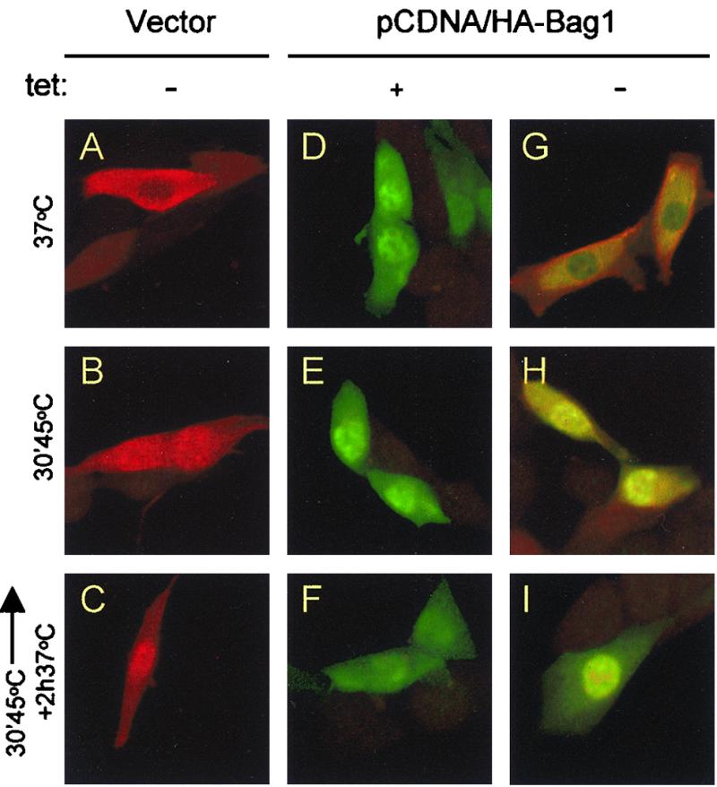
Immunofluorescence analysis of Hsp70 and Bag1 localization. OT70 cells were transiently transfected with pCDNA (vector) or pCDNA/HA-Bag1. Twenty-four hours after transfection the cells were grown in medium with or without tetracycline (tet) for another 24 h. Cells were immunostained after cycloheximide treatment for 30 min (A, D, and G), after heat treatment at 45°C for 30 min (B, E, and H), or after a recovery period for 2 h at 37°C (C, F, and I). All cells were stained with a polyclonal antibody to the heat-inducible Hsp70 and a monoclonal anti-HA tag antibody simultaneously, followed by an incubation with a mixture of tetramethyl rhodamine isocyanate-labeled (red) (Hsp70) and fluorescein isothiocyanate-labeled (green) (HA-Bag1) secondary antibodies. The images were made by confocal microscopy.