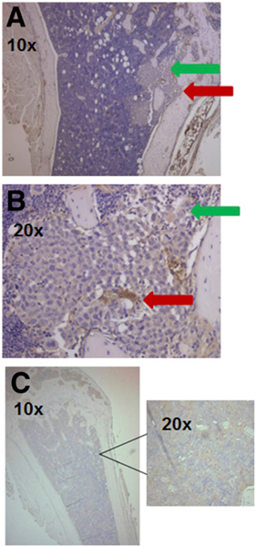
FIGURE 4.
Tumor cells are not α4β1-positive. (A) Histologic sections of mouse leg bones stained with anti–α4 antibody showing either tumor (green arrow) or α4β1-positive HPC (red arrow). (B) Same slide at higher magnification. (C) Histologic section of bone from non–tumor-bearing mouse showing no staining from anti–α4 antibody or at higher magnification.

