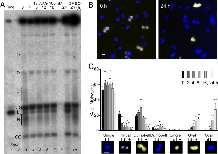
FIG 3.
17-AAG interferes with network remodeling. (A) Southern blot of free minicircles isolated from 100 nM 17-AAG-treated trypanosomes and separated in ethidium agarose. Lane 1, linear minicircle marker; lanes 3 to 7 and 9, increasing duration of 17-AAG; lane 10, controls treated with DMSO for 24 h. Lanes loaded by equal cell equivalents. O, oligomers of two or more interlocked circles; T, theta structures; N/G, mature nicked or gapped daughters; L, linears, N, immature nicked daughters; CC, covalently closed template circles. (B) Kinetoplasts isolated from cells treated for 0 or 24 h with 100 nM 17-AAG and then labeled by TdT with fluorescent dUTP to reveal discontinuities in replicated minicircles (yellow) and stained with DAPI (blue). White bar, 1 μm. (C) Morphology and TdT labeling of networks isolated from trypanosomes treated for increasing intervals with 100 nM 17-AAG. Insets, example images of network morphologies; blue, DAPI; yellow, TdT-incorporated dUTP. White bar, 1 μm. Values are means ± SD from independent experiments; at each point, n = 3 to 9. *, P < 0.05; **, P < 0.01 t test, two-tailed equal variance to 0-h control.

