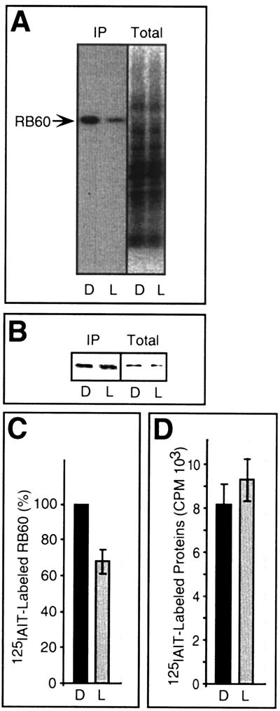
FIG. 5.
In vivo characterization of the redox state of RB60 and total proteins in dark- and light-incubated chloroplasts. (A) Autoradiograph of immunoprecipitation assays of RB60 (IP) conducted with extracts from isolated chloroplasts treated for 10 min in the dark (D) or light (L), followed by [125I]IAIT labeling. Total [125I]IAIT-labeled proteins (without immunoprecipitation) (Total) represent 5% of the amount used for immunoprecipitation assays. (B) The same blot as in panel A, probed with anti-RB60 sera showing equal loading of RB60 protein. (C) Quantification of [125I]IAIT-labeled RB60 (as determined by PhosphorImager analysis and normalized for equal amounts of precipitated RB60 protein) in 10-min light- and dark-treated chloroplasts. Values are means of six independent experiments, and the light value is expressed as percentage of the corresponding dark value (in each experiment designated as 100% of [125I]IAIT-labeled RB60). (D) Quantification of total chloroplast proteins labeled with [125I]IAIT, showing that light does not affect the global protein thiol state in the chloroplast. Each value is an average of three replications.

