. 2021 Oct 4;10:e65672. doi: 10.7554/eLife.65672

Figure 1. Myogenin is required for microtubule-organizing center (MTOC) protein localization to the nuclear envelope.

(A, B) C2C12 cells were differentiated for 1 day and immunostained for the myogenic regulatory factors (MRFs) MyoD or myogenin (Myog) and nesprin-1α (A) or PCM-1 (B). Orange asterisks: MRF+/PCM-1+ nuclei; yellow asterisks: MRF+/nesprin-1α+ nuclei; arrowheads: MRF+/nesprin-1α- nuclei; arrows: MRF-/nesprin-1α+ nuclei. (C) Quantification of (A) and (B). (D) Quantification of MyoD and Myog in relation to nesprin-1α showing that not all nesprin-1α+ nuclei are myogenin+. (E, F) C2C12 myoblasts were transfected with negative control (si-ctrl) or myogenin siRNA (si-Myog) and differentiated for 2 days. Immunostaining (E) and subsequent quantification (F) shows that myogenin depletion affects nuclear envelope localization of PCM-1, PCNT, and AKAP9 but not of nesprin-1α. 95% CI of differences si-Myog vs. si-ctrl = –3.11% to 0.98% (nesprin-1α+), –11.31% to –7.22% (PCM-1+), –7.53% to 3.43% (PCNT+), and –7.52% to –3.42% (AKAP9+). (G, H) C2C12 myoblasts were transfected with si-ctrl or Pcm1 siRNA (si-PCM1) and differentiated for 2 days. PCNT was detected by immunostaining (G) and subsequent quantification (H) showed that PCM-1 depletion reduces PCNT nuclei. 95% CI of differences si-PCM1 vs. si-ctrl = –6.6% to –3.4% (full), –3.78% to –0.59% (partial). Scale bars (A, B, E, G): 20 µm. Data (C, D, F, H) are represented as individual biological replicates (n = 3), together with mean ± SD. ns: p>0.05; *p<0.05; ***p<0.001.

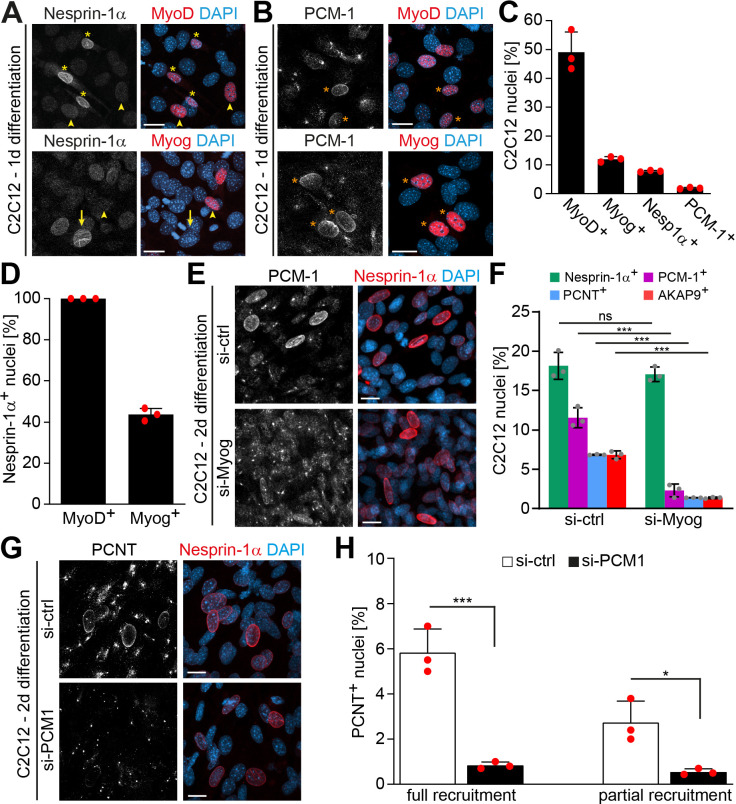
Figure 1—source data 1. Underlying data for graphs in Figure 1C, D and F.

Figure 1.

Figure 1—figure supplement 1. Intermediate stages of microtubule-organizing center (MTOC) protein localization to the nuclear envelope.

Figure 1—figure supplement 1.

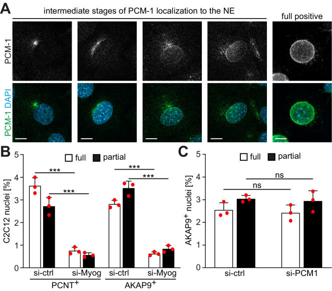
(A) C2C12 cells were differentiated for 24 hr and PCM-1 was detected by immunostaining. Representative images of different intermediate stages of PCM-1 recruitment are shown. Scale bar: 10 µm. (B) A more detailed quantification of PCNT+ and AKAP9+ nuclei in differentiating C2C12 (treated as described in Figure 1F) showing that myogenin depletion affects also partial recruitment of PCNT and AKAP9. (C) C2C12 myoblasts were transfected with si-ctrl or Pcm1 siRNA (si-PCM1) and differentiated for 2 days. AKAP9 was detected by immunostaining and subsequent quantification showed that PCM-1 depletion does not affect AKAP9+ nuclei. Data (B, C) are represented as individual biological replicates, together with mean ± SD. ns: p<0.05; ***p<0.001; n = 3.
Figure 1—figure supplement 2. PCM-1 exhibits a centrosomal localization pattern at nesprin-1+ nuclei in myogenin-depleted C2C12 cells.

Figure 1—figure supplement 2.

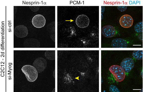
C2C12 myoblasts were transfected with negative control (si-ctrl) or myogenin siRNA (si-Myog) and differentiated for 2 days. In si-ctrl cultures, PCM-1 frequently localizes to the envelope of highly nesprin-1+ nuclei (yellow arrow). In si-Myog cultures, PCM-1 can be found in a centrosomal pattern near nesprin-1+ nuclei (yellow arrowhead). Scale bar: 10 µm.