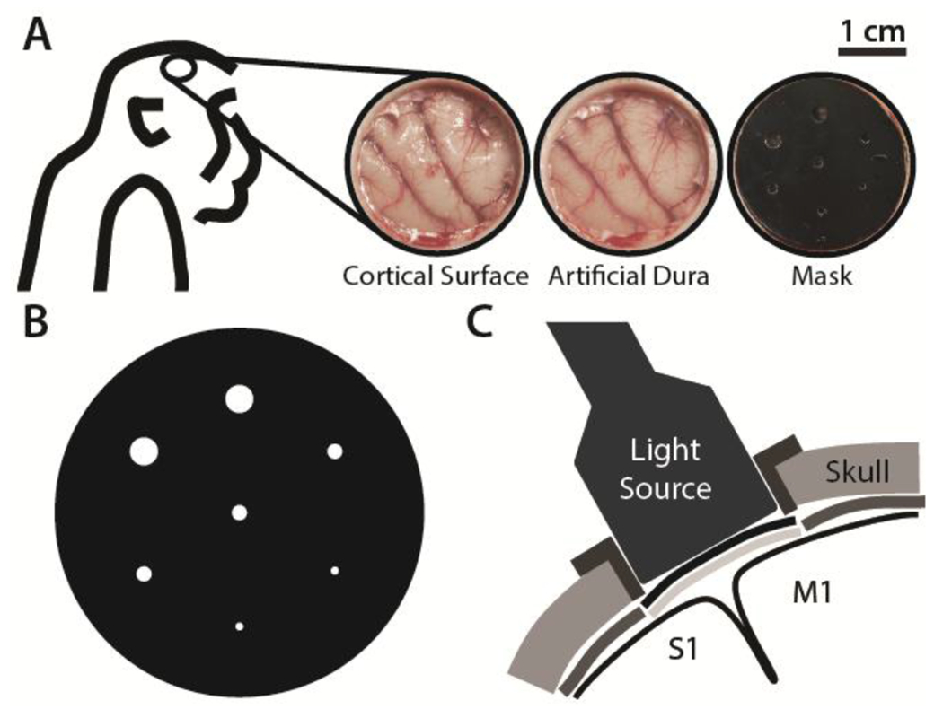
Figure 1. Photothrombotic procedure.

A) Craniotomy over the sensorimotor cortex exposing the cortical surface (left). Transparent artificial dura provides optical access (middle). Opaque mask with apertures for controlled light beam diameters is placed over artificial dura (right). B) Opaque mask schematic showing apertures of diameters 0.5, 1.0, and 2.0 mm. C) Schematic of the light source illuminating primary motor (M1) and somatosensory (S1) cortices through the mask apertures and the transparent artificial dura after intravenous injection of Rose Bengal dye.
