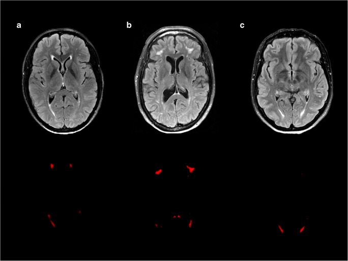
Fig. 3.
An example of white matter hyperintensities (WMH) in all groups of patients. The first row shows the WMH on the FLAIR brain MRI scans. The second row shows the WMH probability maps. a Non-neuropsychiatric systemic lupus erythematosus (non-NPSLE). Female, 37 years old. The MRI scan shows some periventricular and small deep WMH (total WMH volume: 3.04 ml). b Inflammatory neuropsychiatric systemic lupus erythematosus (inflammatory NPSLE). Female, 47 years old. The MRI scan shows deep and confluent WMH (total WMH volume: 18.52 ml) and cerebral atrophy. c Ischemic neuropsychiatric systemic lupus erythematosus (ischemic NPSLE). Female, 47 years old. The MRI scan shows some periventricular and deep WMH (total WMH volume: 3.31 ml)

