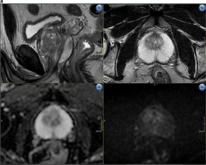
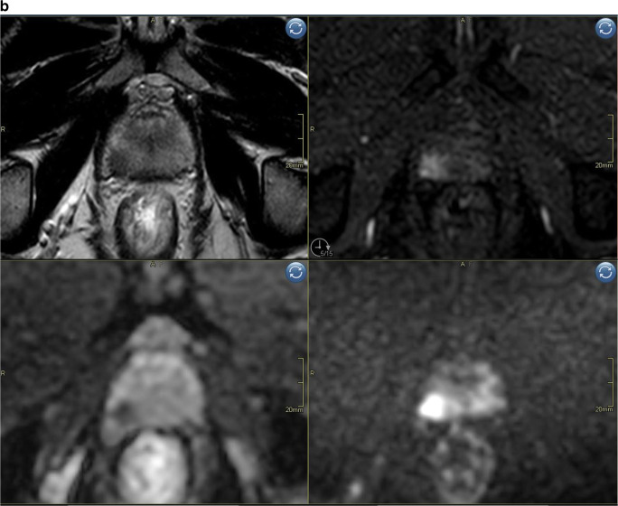
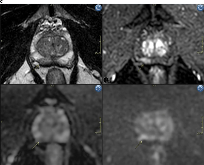
Fig. 3.
a Biparametric MRI example case PIRADS 1 to show image quality. Top left sagittal T2, top right axial T2, bottom left ADC-map, bottom right high b-value (acquired b1500). b Example case PIRADS 4 detected both with bpMRI and mpMRI, no additional value of DCE for tumor detection. Top left axial T2, top right axial DCE, bottom left ADC-map, bottom right DWI b1500. Index lesion in the peripheral zone on the right side with markedly restricted diffusion, early contrast enhancement and size < 1.5 cm. c Example case illustrating a false positive case scored PIRADS 2 (DWI = 2) with bpMRI and PIRADS 4 (DWI = 3; DCE = pos) with mpMRI. Top left axial T2, top right axial DCE, bottom left ADC-map, bottom right high b-value DWI (b1500). Periheral zone index lesion on the right side (yellow arrow)


