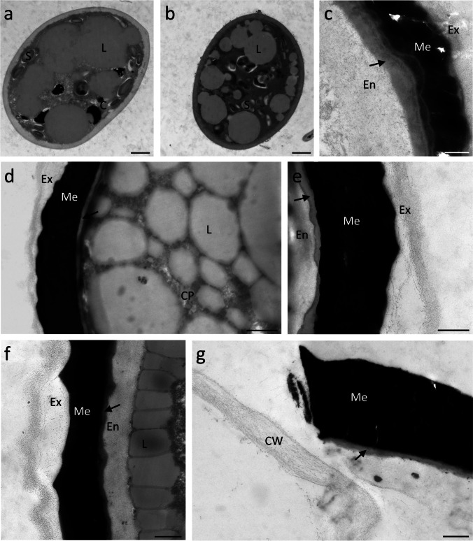
Fig. 3.
Transmission electron micrographs of Mougeotia spp.. M. parvula (a-c), M. disjuncta (d-g). (a) young zygospore with single layered cell wall, (b) maturing zygospore showing the incipient development of a multilayered cell wall, (c) detail view of mature zygospore wall, (d) zygospore with lipid bodies spread through cell lumen, (e) detail view of mature zygospore wall, (f) detail view of zygospore wall with lipid bodies accumulated at the periphery of the cytoplasm, (g) pried open zygospore releasing a newly formed filament. Abbreviations: C chloroplast , CP cytoplasma, L lipid body, S starch grain, En endospore, Ex exospore, Me mesospore, arrow indicates a lipid-like fourth layer. Scale bars (a, b, d) 1 µm, (c, e–g) 500 nm

