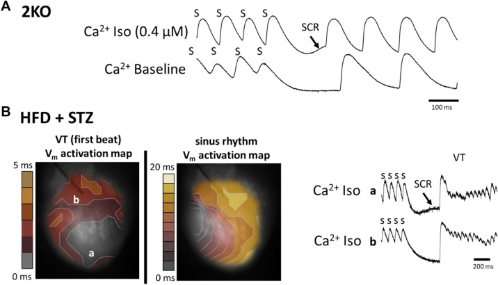
Figure 6.
Examples of arrhythmias induced by isoproterenol (Iso) challenge and spontaneous calcium release (SCR) in Leprdb/db eNOS−/− (2KO) and high fat diet plus low dose STZ-treated (HFD + STZ) mice. (A) Calcium transients with evidence of spontaneous calcium release (SCR, arrow) following the termination of rapid pacing (S) only in the presence of Iso (top trace). (B) Dual voltage-calcium optical mapping results. Activation maps were determined from action potentials measured simultaneously with calcium transients. The site of arrhythmia initiation (VT first beat, left panel B) is associated with the location of SCR (site a), rather than the absence of SCR (site b) following rapid pacing (s), as depicted in the calcium transient traces (right, Panel B). The activation map corresponding to sinus rhythm is shown in the right panel as a reference. Note the difference in time scale between the two maps.

