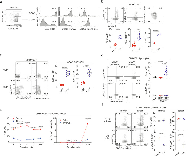
Fig. 1. Peripheral naive CD8+ T cells are phenotypically heterogeneous.
a Ly6C, CD183, and CD103 expression on B6 CD44lo and CD44hi CD8+ T cells. b Percentage of Ly6C+, CD183+, and CD103− cells in CD5lo and CD5hi B6 naive CD8+ T cells (n = 12 mice for Ly6C+ and CD183+, n = 5 mice for CD103−). c Percentage of CD183+ and CD103− cells in Ly6C− and Ly6C+ CD5hi B6 naive CD8+ T cells (n = 12 mice for CD183+, n = 7 mice for CD103−). d Percentage of CD183+ and Ly6C+ cells in CD24hi, CD24lo CD5lo, and CD24lo CD5hi B6 CD4−CD8+ thymocytes (n = 12 mice for Ly6C+, n = 6 mice for CD183+). e Splenic and thymic Ly6C+ B6 naive CD8+ T cell changes over time after birth (n = 3 mice for each group). f Splenic and thymic Ly6C+ B6 naive CD8+ T cells in young and old mice (n = 3 mice for each group). Statistical significance was confirmed by two-tailed unpaired Student’s t-test. Results are shown as mean ± SD. Data representative of 2–3 independent experiments. Source data are provided as a Source data file.

