
Figure 4.
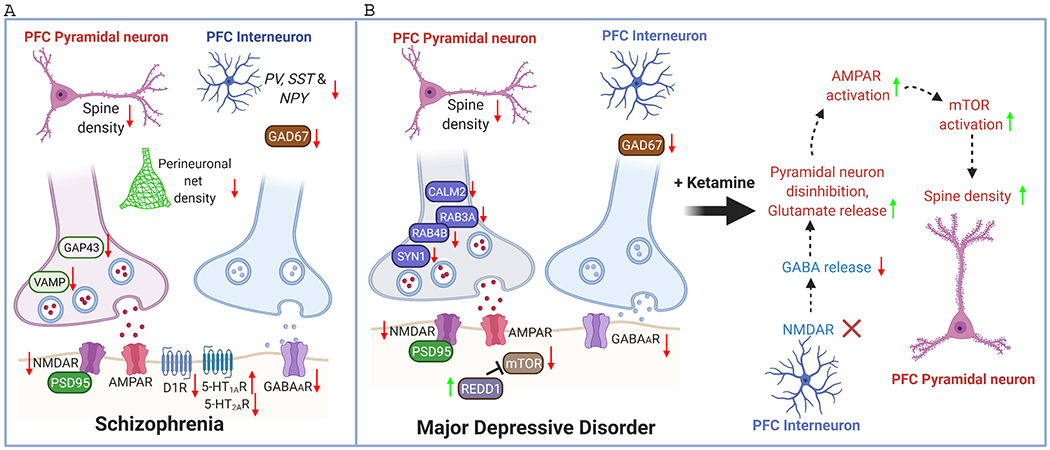
Schematic diagram illustrating the synaptic changes in PFC of schizophrenia (SZ) and major depressive disorder (MDD). NMDAR hypofunction, GABA deficiency, dopamine hypoactivity, serotonin dysregulation, and dendritic spine loss are major aberrations in SZ PFC. The reduced density of perineuronal nets (PNN) may contriubute to the destabilization of synapses. Similar reduction on NMDAR, GABA system and spine density is found in MDD PFC. Some presynaptic molecules involved in transmitter release, including CALM2, SYN1, RAB3A, RAB4B, are also diminished in MDD PFC. In addition, mTOR signaling is decreased in MDD PFC because of the elevated expression of the endogenous inhibitor REDD1. The fast-acting antidepressant ketamine primarily blocks NMDARs on PFC interneurons, leading to the disinhibition of PFC pyramidal neurons and the increase of mTORC signaling, resulting in the restoration of dendritic spines in MDD PFC.