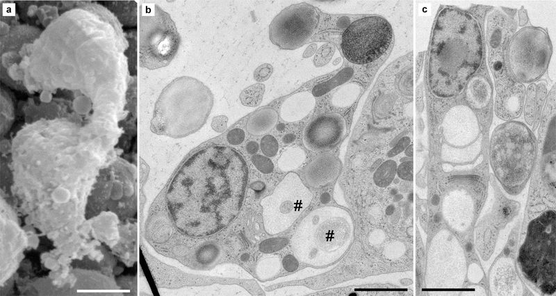
Fig. 4.
Ultrastructure of lipophil cells in H. hongkongensis (H13). a At the SEM, lipophil cells are at times seen bulging from the lower epithelium into the fiber cell layer, often in close vicinity to fiber cells. Their shapes are usually elongated (in the lower epithelium, where they are interspersed between lower epithelial cells (c) or more complex depending on their cellular environment (b) and position in the animal. In all cases, they exhibit large, heterogeneous vacuoles— rather clear for those close to the trans-Golgi network, otherwise with various electron densities (i.e., affinity to osmium) or structural complexity, as some of the vacuoles contain membranous material (#). Their most basal vacuole (matte-finished sphere—asterisk), abutting the lower surface, is often electron-dense but sometimes also clear. Scale bar 2 μm in a; 1.5 μm in b, c

