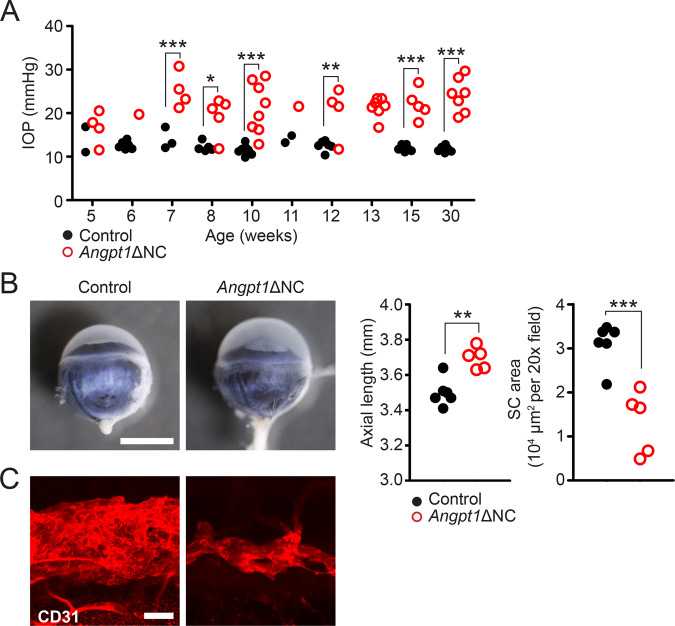
Fig. 2. Angpt1ΔNC mice exhibit ocular hypertension and hypomorphic Schlemm’s canal development.
A Compared to control littermates, rebound tonometry revealed elevated intraocular pressure in Angpt1ΔNC mice beginning at 7 weeks of age. Each group was measured at only a single timepoint, except for one group measured at 10 and 30 weeks of age. Each datapoint represents the average of left and right eyes from a single animal. Genotype p < 0.0001 as determined by two-way ANOVA, Dfgenotype = 1, Fgenotype = 95.67. *p ≤ 0.05, **p ≤ 0.01, ***p ≤ 0.001 as determined by Bonferroni posttests. Lacking littermate controls, 13-week timepoint was excluded from statistical analysis. n = 36 (control) and 45 (Angpt1ΔNC) animals from eight independent litters. B By 15 weeks, increased axial length was apparent in enucleated eye globes of Angpt1ΔNC mice. n = 6 (control), 5 (Angpt1ΔNC), p = 0.0013. C Compared to littermate controls, whole-mount confocal microscopy using anti-CD31 antibody revealed reduced Schlemm’s canal area in Angpt1ΔNC mice, suggesting that ocular hypertension was due to insufficient aqueous humor outflow. n = 6 (control), 5 (Angpt1ΔNC), p = 0.0008. **p ≤ 0.01, ***p ≤ 0.001 as determined by two-tailed Student’s t-test. Df = 9. Scale bars represent 2 mm (B) and 50 μm (C). 20× fields used for quantification in (B) represent an area of 65,025 μm2.

