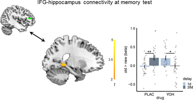
Fig. 3. Noradrenergic stimulation reverses the time-dependent changes in IFG-hippocampus functional connectivity.
Psychophysiological interaction analysis indicated that while the connectivity between the inferior frontal gyrus (IFG) and hippocampus increased in the placebo (PLAC) group from 28d relative to 1d (p = 0.004 two-tailed Welch’s t-test), there was even a decrease in IFG-hippocampus connectivity at 28d compared to memory testing after 1d in the yohimbine (YOH) group (p = 0.020, two-tailed Welch’s t-test; drug × delay, SVC peak-level: x = −22, y = −40, z = −2, p(FWE) = 0.009, mixed ANOVA; n = 104 participants). Bonferroni correction was applied for the number of regions of interest in each analysis. The seed-region in the IFG (green), retrieved from the drug × delay interaction of the univariate analysis (peak: x = −50, y = 34, z = 12; k = 62), and the significant cluster in the hippocampus (orange) are superimposed on sagittal slices of T1-weighted template images. Distribution of beta-values for the significant cluster is presented for the contrast old > new. Bars represent mean ± SEM. Source data are provided as Source data file. *p < 0.050, **p < 0.010.

