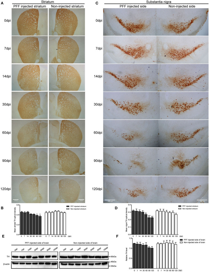
Figure 5.
Dopaminergic neuronal density decreased in the striatum and SN following PFF injection. (A,B) TH-positive dopaminergic nerve terminal density in the striatum decreased with time in PFF-injected and noninjected sides, which are shown in the DAB image (A) and its quantification data (B) (χ2 = 41.72, p < 0.0001, df = 13). (C,D) TH-positive dopaminergic neurons decreased with time in the bilateral SN, especially PFF-injected SN, which is shown in the DAB image (C) and quantification (D) (χ2 = 33.64, p = 0.0014, df = 13). (E) The western blotting analysis of the whole brain lysate of the PFF-injected side of the brain and the noninjected side of the brain at various time points, using antibodies against TH. Original blots are presented in Supplementary Figure 7. (F) The relative TH expression level in the PFF-injected side of the brain (χ2 = 12.00, p = 0.5278, df = 13). Bars indicate SEM, n = 3–4/time point. Significance was measured by a comparison of 0 dpi to each time point by post hoc analysis (the Kruskal–Wallis test, *p < 0.05, ***p < 0.001). Scale bar, 500 μm.

