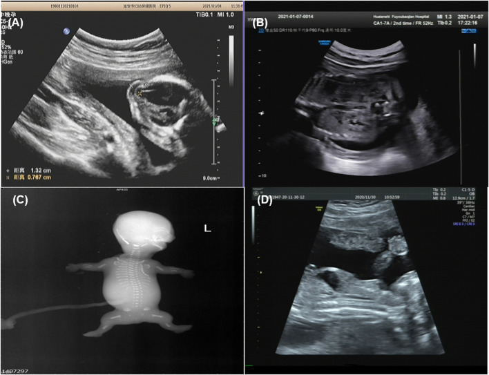
FIGURE 2.
Imaging examination of ciliary diseases {Family 1 [(A): case 2], Family 3 [(B): case 5], Family 4 [(C): case 6], Family 5 [(D): case 8], occipital encephalocele (A), increased echogenicity in both renal cortexes (B), fetal X-ray showing bilateral shortened curved femora and disproportionately shortened tibiae (C), bone dysplasia and narrow chest (D)}.

