FIGURE 2.
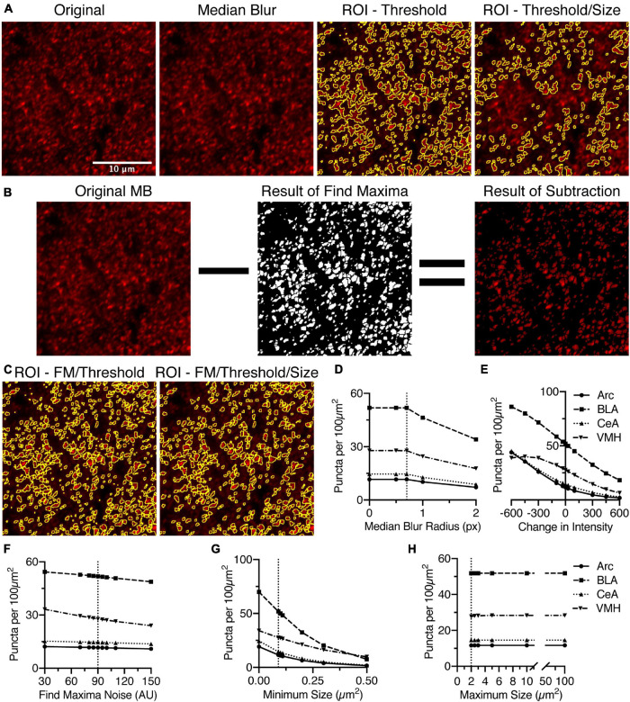
Identification of puncta in densely labeled images requires a number of settings, but is most affected by the threshold intensity and the minimum size. (A) Anti-bassoon staining in BLA is densely labeled but clearly denotes puncta. Median blur with a sub-pixel radius removes noise without noticeably affecting the puncta. Identifying puncta using analyze particles above a minimum threshold, removing small and large puncta, leads to significant loss of most of the stained puncta. (B) Alternatively, the “Find Maxima” function (FM) identifies borders between puncta above the threshold using the segmented particles setting. This creates a black and white image, which with “Image Math,” can be used to remove pixels below threshold and create borders around closely spaced puncta. (C) Anti-bassoon puncta identified using thresholding and FM is further refined by using the analyze particles function to remove puncta outside a minimum and maximum size range (Size). Number of puncta identified in anti-bassoon staining in four brain regions is maximally, and universally negatively correlated with changes in intensity threshold (E) and a minimum size (G). Sub-pixel median blur (D), changes in the noise setting for FM (F), and maximum size (H) have comparatively little effect on the number of identified puncta. Vertical dotted lines denote the original starting setting used for analysis. Scale bar denoted in panel (A) is 10 μm for all images.

