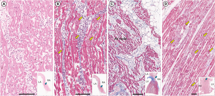
Fig. 1. Histopathology of the heart. (A) Hematoxylin and eosin stains of atrial septum shows massive inflammatory infiltration with neutrophil predominance. (B) The myocytes often show contraction band necrosis (yellow arrows), which were highlighted by Masson's trichrome staining. (C) The atrioventricular node area shows extension of atrial myocarditis to the superficial layer of the node. (D) The ventricular myocardium is free of inflammatory infiltrates, but there are multiple large foci of contraction band necrosis (yellow arrows) particularly in the left ventricular wall and the ventricular septum. Bars represent 100 µm. The blue arrows in insets show where the section was taken from the low magnification views. Hematoxylin and eosin stain was used for the specimen shown in (A) and Masson's trichrome stain was used for the specimen shown in (B-D).
RA = right atrium, LA = left atrium, RV = right ventricle, LV = left ventricle.

