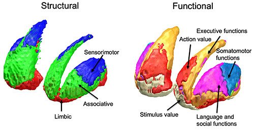
Figure 3.

Striatal topography according to structural and functional connectivity. Right: Striatal functional territories obtained by diffusion tractography141 are arranged along the ventro-dorsal and rostro-caudal axes. The ventral striatum (red) is mainly connected to ventromedial prefrontal cortices, the antero-dorsal territory (green) or the central striatum has stronger connectivity with prefrontal areas related to cognition, the postero-dorsal territory (blue) or the posterior striatum is mainly connected to sensorimotor areas. Left: Striatal territories defined using resting-state functional MRI, classified according corresponding cortical networks as follows: limbic (cream), default mode (red), frontoparietal (orange), somatomotor (light blue), dorsal attention (green), ventral attention (purple) and visual (violet).110 Behavioral labeling follows task-based activation of selected striatal territories.115
