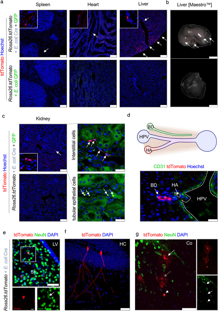
FIGURE 5.

Bacterial OMVs transfer functional Cre mRNA to multiple organs. (a–g) Representative data derived from different organ tissue of Rosa26.tdTomato mice treated with either E. coli Cre plus E. coli GFP (n = 9) or with E. coli GFP only (n = 5). Experiments were repeated 3 times with similar results. (a) Representative confocal images of different organ cross sections of spleen, heart and liver showing tdTomato‐positive cells (red). Nuclei counterstaining with Hoechst (blue). Scale bar: 100 μm. (b) Representative Maestro ex vivo images of the liver of Rosa26.tdTomato mice treated with E. coli Cre (plus E. coli GFP) or E. coli GFP as control analyzed via multi‐spectral separation. Scale bar: 50 mm. (c) Representative confocal images of kidney cross‐sections showing tdTomato‐positive cells (red). Tubular epithelium shown in green. Nuclei counterstaining with Hoechst (blue). Scale bar: 100 μm. (d) Representative pictures of tdTomato positive biliary epithelial cells in liver cross sections. Scale bar: 50 μM. Arrows point towards tdTomato‐positive cells. HPV: hepatic portal vein; HA: hepatic artery; BD: bile duct. Scale bar: 25 μm. (e‐g) Representative confocal images of immunohistochemical staining of brain cross‐section showing tdTomato‐positive cells (red) and staining against NeuN (e,g) (Green, Neuron). Nuclei counterstaining with DAPI (blue). Scale bar: 50 μm (Upper image), 10 μm (Lower image). LV: lateral ventricles; St: striatum; HC: hippocampus; Co: cortex
