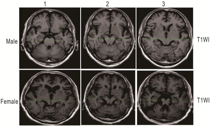
FIGURE 1.
As a measure of external cerebral atrophy, SFR, the average of the maximum width of the two Sylvian fissures on the section showing them at their widest, divided by the transpineal coronal inner table diameter. Illustrative measurement of brain MRI images showing green lines in female and male patients, respectively. Measurement values are automatically generated by imaging software. Upper: A 73-year-old man with SFR 0.051: MoCA score: 20 on T1WI. Lower: A 76-year-old woman with SFR 0.068: MoCA score: 14 on T1WI. T1WI, T1 weighted image.

