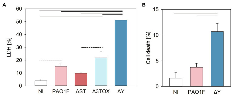
Figure 5.
Cytotoxicity measured by lactate dehydrogenase (LDH) release (A) or fluorochrome 7-amino actinomycin D (7-AAD) detection of cell damage (B) in human bronchial NCI-H292 cells infected by P. aeruginosa strains for 3h at MOI 20. (A) Cytotoxicity was assessed based on the amount LDH released by epithelial cells into the culture supernatant measured as detailed in the Materials and Methods (determined as % of total cell LDH). Mean values and SD were calculated from three independent measurements taken using the CytoTox 96® Non-Radioactive Cytotoxicity Assay kit. One way ANOVA analysis with subsequent pairwise comparison (Turkey method) revealed significant differences between pairs connected by solid lines or dashed lines indicating value of p<0.001 or <0.005, respectively. (B) 7-AAD-mediated detection of cell damage. Cells were stained with 7-AAD as described in Materials and Methods and analyzed with a cytoFLEX Beckman Coulter Flow Cytometer and FlowJo software. Mean values and SD correspond to three independent experiments with samples measured in duplicate. Experiments were performed on NCI-H292 that had undergone between 8 and 20 passages. Pseudomonas aeruginosa strains expressed the following T3SS effectors: PAO1F, wild-type expressed ExoS, ExoT, and ExoY; ΔST expressed only ExoY; Δ3TOX, expressed none of the T3SS effectors; and ΔY expressed ExoS and ExoT. One way ANOVA analysis revealed significant differences (p<0.001) between PAO1F and ΔY.

