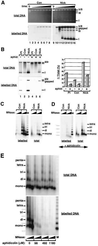
FIG. 2.
Single-strand breaks trigger chromatin assembly in the absence of extensive DNA synthesis. (A) Supercoiling analysis of chromatin assembly on DNA substrates containing site-specific single-strand breaks after incubation in a Drosophila cell-free system. Incubation times were 1 min (lanes 2 and 10), 5 min (lanes 3 and 11), 15 min (lanes 4 and 12), 45 min (lanes 5 and 13), 90 min (lanes 6 and 14), 180 min (lanes 7 and 15), and 360 min (lanes 8 and 16). Lanes 1 and 9 contain untreated Con and nick DNA, respectively. The migration of relaxed/nicked circular DNA (Ir/II), linear DNA (III), supercoiled DNA (I), and labeled gapped circular molecules is indicated. (B) Supercoiling analysis of chromatin assembly using DNA substrates containing site-specific DNA lesions after incubation in a Drosophila cell-free system for 6 h in the presence or absence of aphidicolin (590 μM). The graph shows the quantification of the number of extensively supercoiled topoisomers (I) relative to the total topoisomer population as a percentage. The migration of relaxed/nicked circular DNA (Ir/II), linear DNA (III), supercoiled DNA (I), and labeled gapped circular molecules is indicated. (C) MNase digestion analysis of nucleosomal arrays formed on nick DNA after 6 h incubation in a Drosophila cell-free system. Two digestions with increasing amounts of MNase are shown for each reaction. The regularly spaced DNA bands corresponding to mono-, di-, tri-, and tetranucleosomal DNA are indicated. A 123-bp ladder was used as a molecular weight marker (M). (D) Effect of aphidicolin (590 μM) on nucleosomal arrays formed using nick DNA in a 6-h reaction with the Drosophila cell-free system (MNase digestion analysis was performed under conditions identical to those used for panel C). (E) Formation of nucleosomal arrays using nick DNA in a 6-h reaction with the Drosophila cell-free system in the presence of increasing amounts of aphidicolin. The distinct pattern of MNase digestion products seen in the presence of aphidicolin on labeled Nick DNA molecules in panels D and E is indicated by dashed lines. This pattern is thought to result from the assembly of nucleosomal arrays adjacent to gaps in the damaged DNA strand.

