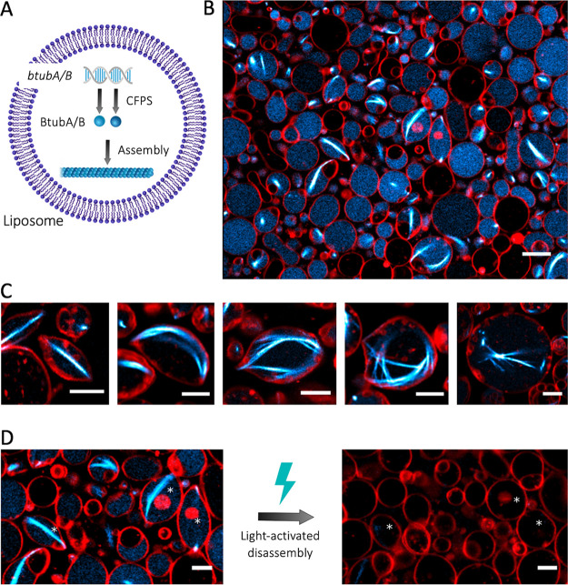
Figure 4.
CFPS and the assembly of bMTs inside liposomes. (A) Schematic of liposome-compartmentalized gene expression and synthesis of BtubA/B that self-organize into bMTs. (B) Fluorescence confocal microscopy images of liposomes (red, DHPE-TexasRed) with encapsulated Atto488-BtubA/B (100 nM, cyan) after 4 h of CFPS reaction. In situ synthesized bMTs (cyan) are visible in several liposomes. Scale bar: 10 μm. (C) Examples of different bMT cytoskeletal structures showing clear membrane deformation. Samples were observed after 5 h of incubation at 37 °C. Scale bars: 5 μm. (D) Breaking of bMTs and relaxation of liposomes into a spherical shape was triggered by the exposure of samples to a high laser intensity. Asterisks indicate liposomes whose shape was modified by the light-activated disassembly of bMTs. Scale bars: 5 μm.

