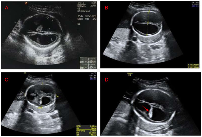
Figure 2.
Transabdominal view of the fetal brain. (A) Ultrasound image of the first hydrocephalus fetus. The width of the left ventricle was 2.05 cm, the width of the right ventricle was 2.65 cm and the cerebral cortex became thinner. (B) Ultrasound image of the second hydrocephalus fetus. The width of the left ventricles was 2.43 cm, the width of the right ventricle was 2.37 cm and the cerebral cortex became thinner. (C) Ultrasound image of the second hydrocephalus fetus. The biparietal diameter of the fetus was 6.06 cm, the occipitofrontal diameter was 7.73 cm and the head circumference was 22.55 cm. (D) Ultrasound image of the second hydrocephalus fetus. Hyperechoic choroid hanging sign (arrow), the bilateral lateral ventricles were expanded and the cerebral cortex became thinner.

