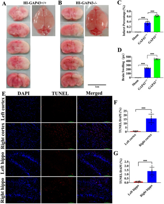
Fig. 4.

Outcomes of GAP43 knockout on cerebral damage and cell survival in HI rats. a, b The gross anatomical characteristic of rat brain surface and TTC staining slices after HI in the GAP43+/+ and GAP43−/− groups, scale bar = 1 cm. c Comparison of brain infarction ratio among the sham, GAP43+/+, GAP43−/− groups. d Brain swelling comparison in the above-mentioned three groups. e TUNEL/DAPI staining images of cortex and hippocampus in the GAP43−/− rats, scale bar = 100 μm. f Cell apoptosis rate of the left and right cortex in the GAP43−/− rats. g Cell apoptosis rate of the left and right hippocampus in the GAP43−/− rats. HI hypoxia ischemia, GAP43+/+ wild-type rat, GAP43−/− GAP43 knockout. ***p < 0.001, n = 5/group
