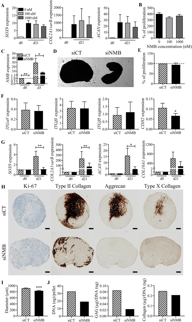
Fig. 5.
Role of NMB during chondrogenesis. A Fold change expression of chondrocyte markers after 21 days of chondrogenic differentiation of BM-MSCs with different concentrations of recombinant NMB. Results are expressed as mean fold change ± SEM (n = 3 biological replicates). B Proliferation of BM-MSCs cultured with different concentrations of recombinant NMB after 5 days of culture. Results are expressed as the mean percentage ± sem (n = 4 biological replicates). C Fold change expression of NMB in BM-MSCs at day 3 after transfection with siRNA control (siCT) or siRNA against NMB (siNMB) (normalized to NMB expression at d0). D Representative picture of micropellets after 3 days of chondrogenic differentiation of BM-MSCs transfected with siCT or siNMB. E Proliferation of BM-MSCs transfected with siCT or siNMB after 3 days of culture in monolayer. Results are expressed as mean percentage ± sem (n = 4 biological replicates). F Relative expression of adhesion molecules in BM-MSCs transfected with siCT or siNMB after 3 days of chondrogenic differentiation. G Fold change expression of chondrocyte markers in BM-MSCs transfected with siCT or siNMB at day 21 of chondrogenesis. Results are expressed as mean ± sem (n = 8 biological replicates). *: p ≤ 0.05, **: p ≤ 0.01. H Immunohistological staining of Ki-67, type II Collagen, Aggrecan and type X Collagen in a micropellet representative of BM-MSCs transfected with siCT or siNMB at day 21 of chondrogenesis (scale bar = 100 µm). I Average diameter of micropellets from BM-MSCs transfected with siCT or siNMB at day 21 of chondrogenesis (n = 15). J Amounts of total DNA, GAG and collagen contained in micropellets of BM-MSCs transfected with siCT or siNMB at day 21 of chondrogenesis (pool of 5 pellets)

