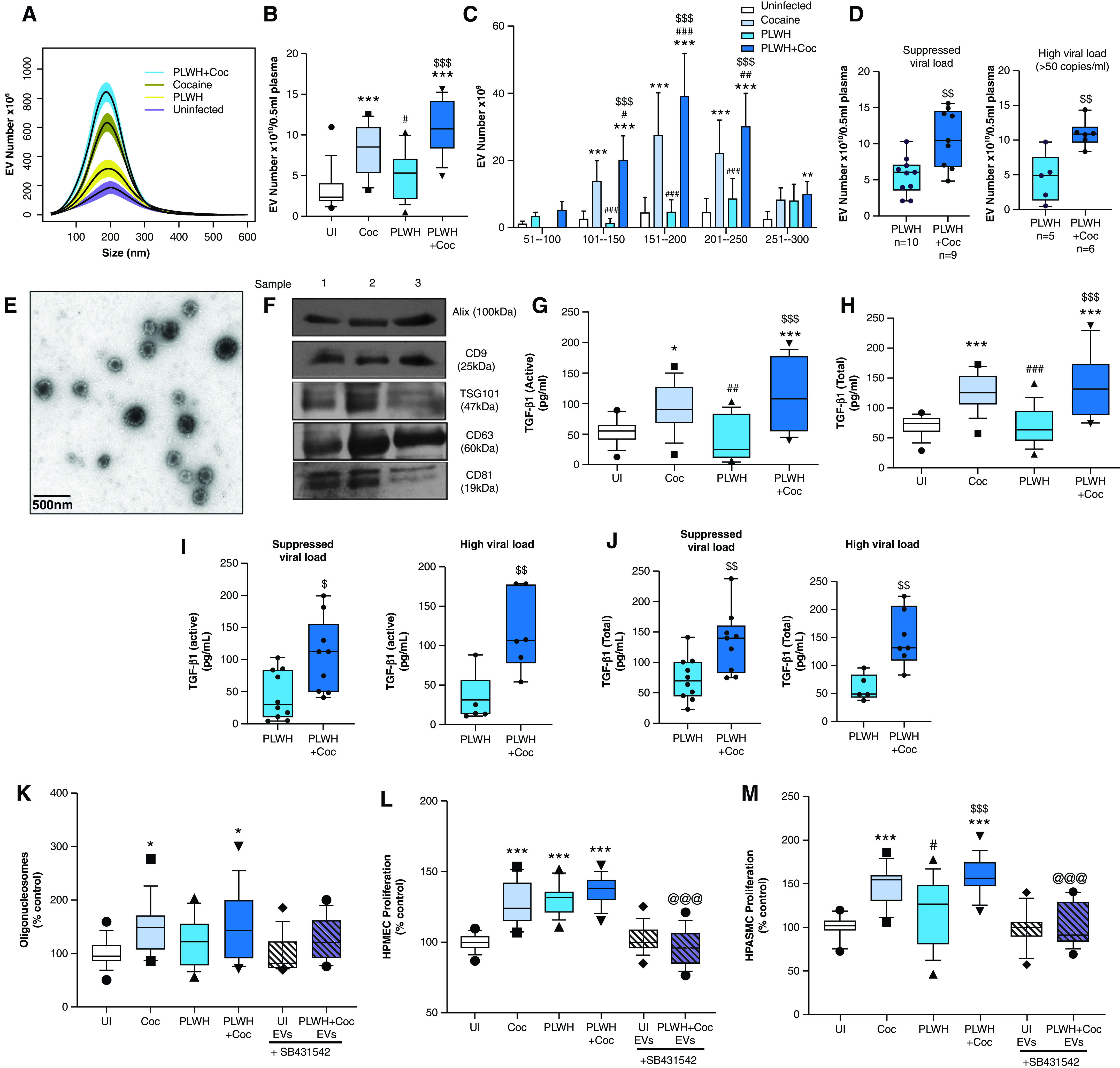
Figure 1.

Analysis of plasma-derived extracellular vesicles (PEVs) from those with human immunodeficiency virus (HIV) and/or a history of cocaine use. Extracellular vesicles (EVs) were isolated from 0.5 milliliter (ml) of EDTA plasma from HIV-negative individuals without a history of cocaine use (UI), HIV-negative individuals with a history of cocaine use (Coc), people living with HIV (PLWH) without a history of cocaine use, and PLWH with a history of cocaine use (PLWH + Coc) (n = 15/group) by using an exoEasy kit. (A–D) Particles were counted and characterized for size distribution by using nanoparticle tracking analysis. Data represent the number of EVs/ml of a final EV suspension obtained from 0.5 ml of EDTA plasma by using an exoEasy kit. The high viral load group included PLWH with ⩾50 viral RNA copies of per ml of plasma. (E–F) Characterization of PEVs by using transmission electron microscopy (TEM) and Western blotting analysis. Representative TEM image of PEVs at 5,000× magnification (E) and Western blot of PEV protein extract from three different subjects for exosomal markers (F). Scale bar, 500 nm. (G–J) PEVs were analyzed for active (G and I) and total (H and J) TGF-β1 (transforming growth factor-β1) levels by using an ELISA. Analysis of (K) endothelial apoptosis and (L) proliferation on treatment with PEVs from HIV-negative individuals without a history of cocaine use, HIV-negative individuals with a history of cocaine use, PLWH, and PLWH + Coc (n = 15/group). Human pulmonary microvascular endothelial cells (HPMECs) were plated in a 96-well plate and were serum-starved after 24 hours in 0.5% serum–containing media, which was followed by the addition of 3 μg of PEVs/well. (K) Analysis of apoptosis by using a cell-death ELISA after 24 hours and (L) analysis of cell proliferation by using a cell proliferation assay after 48 hours. (M) Analysis of human pulmonary arterial smooth muscle cell (HPASMC) proliferation on treatment with PEVs. After 72 hours of plating in a 96-well plate, cells were serum starved for 48 hours, which followed by the addition of 3 μg of PEVs/well. A MTS cell proliferation assay was performed at 48 hours after treatment. Cell proliferation and apoptosis assays were also performed on cells treated with PLWH + Coc PEVs in the presence of 10μM SB431542 (TGFβ-R1 inhibitor [In.]). Boxes represent the median and interquartile range (IQR), and whiskers show the 10th to 90th percentiles. *P < 0.05, **P < 0.01, and ***P < 0.001 versus HIV-negative individuals without a history of cocaine use; #P < 0.05, ##P < 0.01, and ###P < 0.001 versus HIV-negative individuals with a history of cocaine use; $P < 0.05, $$P < 0.01, and $$$P < 0.001 versus PLWH; and @@@P < 0.001 versus PLWH + Coc without TGFβ-R1 In.
