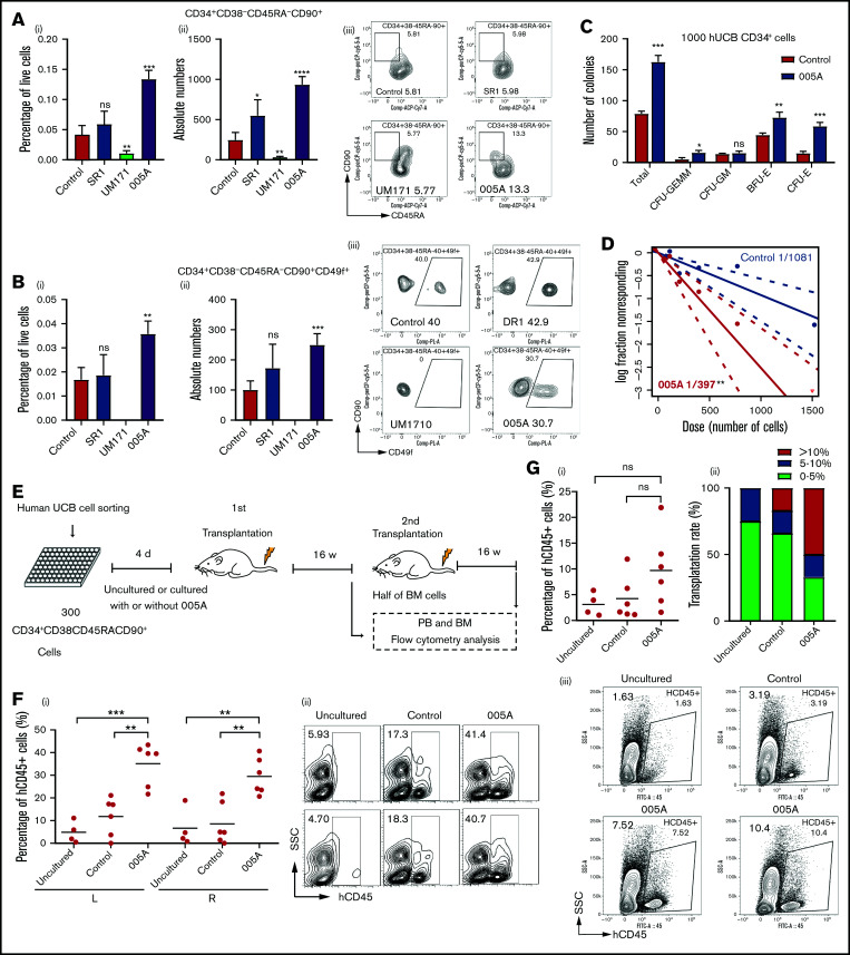
Figure 3.
005A treatment retained the stemness of human primary HSCs. (A) The percentage (i) of live cells and absolute numbers (ii) per well of human CD34+CD38−CD45RA−CD90+ cells were measured (n = 4). Representative images (iii) of the flow cytometry data of CD34+CD38−CD45RA−CD90+ cell population and percentage in CD34+CD38− cells. (B) The percentage (i) of live cells and absolute numbers (ii) per well of human CD34+CD38−CD45RA−CD90+CD49f+ cells were measured (n = 4). Representative images (iii) of the flow cytometry data of CD34+CD38−CD45RA−CD90+CD49f+ cell population and percentage in CD34+CD38−CD45RA−CD90+ cells; 1 × 10⁵ CD34+ UBCs were planted in 24-well plate, and cell populations were analyzed by flow cytometry after 7-day treatment with vehicle, SR1(1 µmol/L), UM171 (35 nmol/L), or 005A (20 nmol/mL) in panels A and B. (C) CFU statistical analysis of colony formation data for cells treated with or without 005A for 7 days (n = 3). GEMM, colony-forming unit-granulocyte, erythroid, macrophage, megakaryocyte; CFU-GM, colony-forming unit-granulocyte, macrophage; BFU-E, Burst-forming unit-erythroid; CFU-E, colony-forming unit-erythroid. (D) Frequencies of CAFCs of vehicle and 005A-treated human CD34+ cells (n = 10). Doses of 1500, 750, 375, 186, and 92 cells per well were used. The experiment was repeated 3 times. (E) An overview of the experimental setup used to perform xenotransplantations in immunodeficient NOG mice. (F) Levels of human engraftment (i) and representative images of the flow cytometry data (ii) in intrafemoral injected (R, right) or none injected (L, left) bone marrow from primary recipients of human CD34+CD38−CD45RA−CD90+ cells that were directly transplanted (uncultured) or cultured with or without 005A (n = 4 for uncultured group, n = 6 for control group and 005A group). Each symbol represents the results from an individual mouse. (G) Levels of human engraftment (i) of the secondary NOG recipients and chimerism were assessed after 4 months. (ii) Transplantation rate of mice at different engraftment levels (<5%; 5%−10%; >10%) in each group are shown. n = 4 for uncultured group, n = 6 for control group and 005A group. (iii) Representative fluorescence-activated cell sorter profiles. All data represent means ± SD. Compared with control unless specified: *P < .05, **P < .01, ***P < .001 by 2-tailed unpaired t test.

