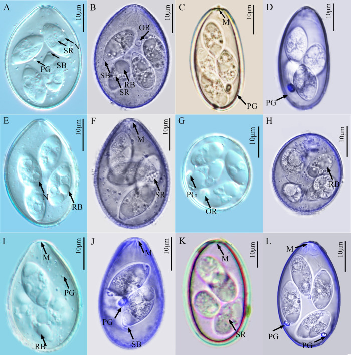
Figure 1.
Photomicrographs of the sporulated oocysts of six Eimeria species collected from forest musk deer. A, B. Eimeria aquae n. sp.; C, D. Eimeria dolichocystis n. sp.; E, F. Eimeria fengxianensis n. sp.; G, H. Eimeria helini n. sp.; I, J. Eimeria kaii n. sp.; K, L. Eimeria oocylindrica n. sp.; Figs. A, E, G, I were obtained by differential interference contrast microscopy; Figs. B, D, F, H, J, L were obtained by confocal laser scanning microscopy; Figs. C, K were obtained under standard light microscopy under oil immersion. Abbreviations: M, micropyle; PG, polar granule; OR, oocyst residuum; RB, refractile body in sporozoite; SB, Stieda body on sporocyst; SR, sporocyst residuum; N, nucleus of SZ.

