
Fig. 2.
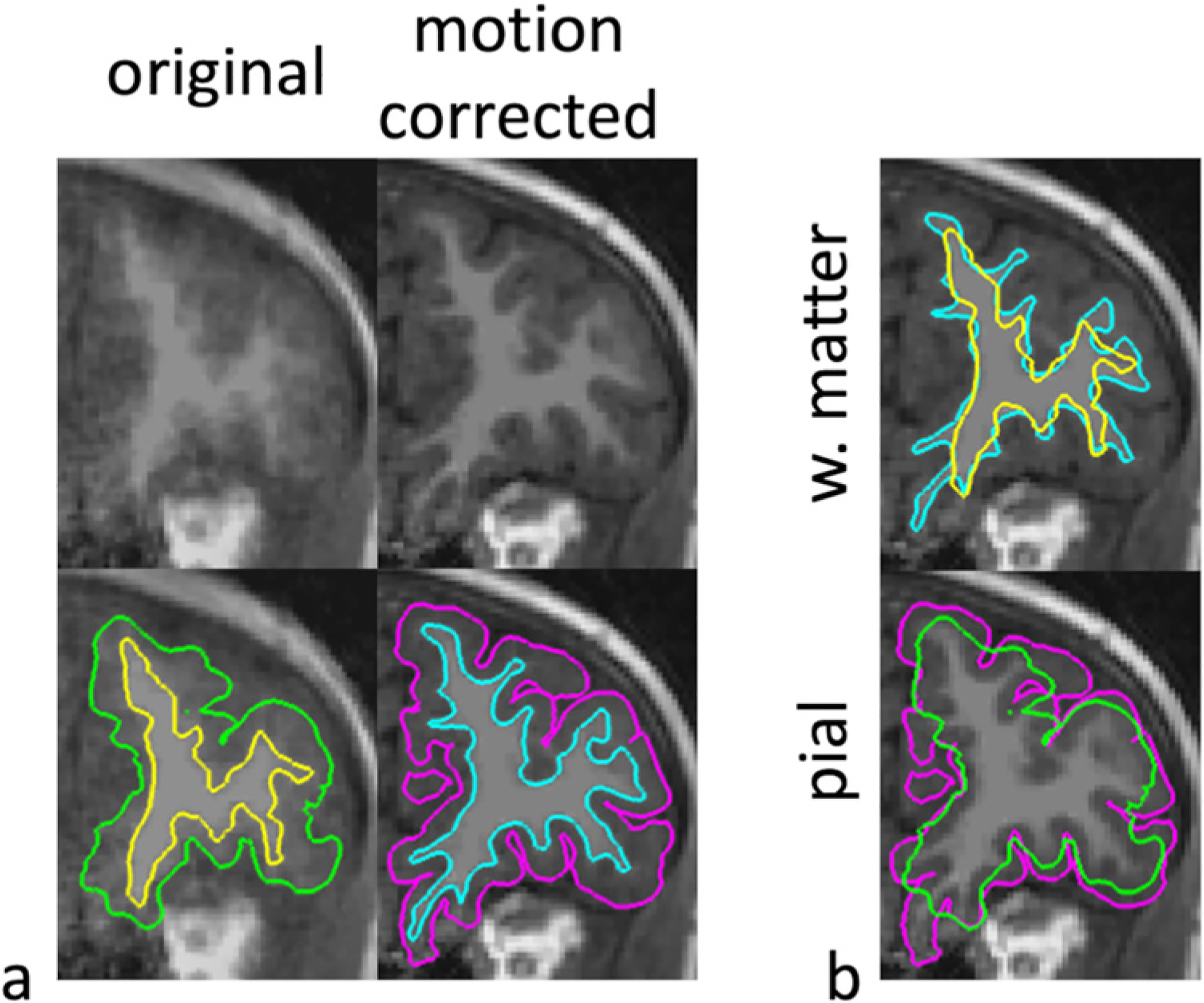
The resulting cortical segmentations (pial and white matter surfaces) from the T1-weighted image in Fig. 1 are shown for MPnRAGE without (No MC) and with (MC) motion correction. The white matter (aqua and yellow) and pial (magenta and green) are shown with and without motion correction respectively in (b).