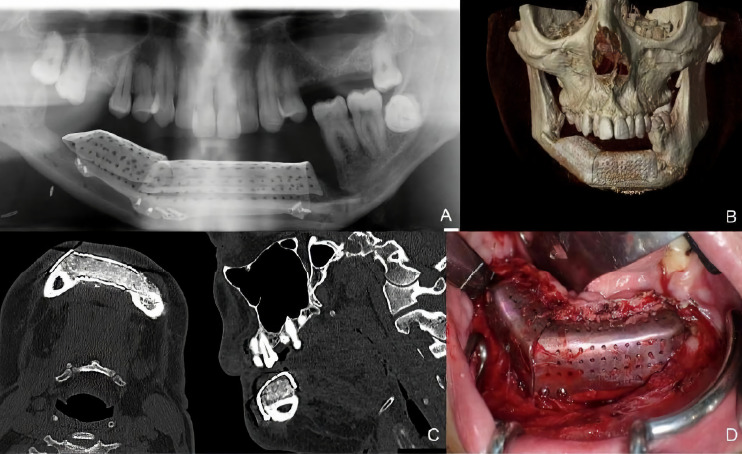
Figure 7.
(A) Panoramic radiograph showing a bone gap between the iliac crest graft and the remnant mandible mesially to the lower molar due to the angular shape of the titanium mesh at this level. CT Scan (B) with three-dimensional reconstruction of the mandible. CT Scan demonstrating the stability of the transverse dimension of the fibula with respect to the remnant mandible. (C) The three-dimensional preservation of the iliac crest graft with CAD/CAM mesh makes it possible to double the height of the fibula. (D) Intraoral approach showing the three-dimensional preservation of the customized mesh.

