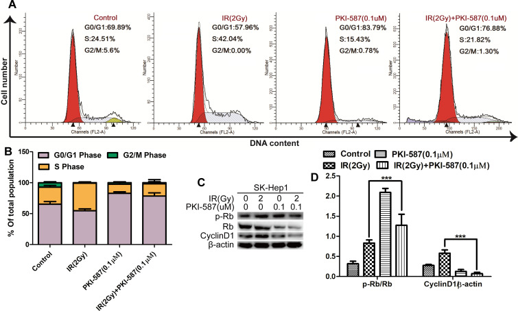
Fig 4. PKI-587 combined with IR promoted G0/G1 phase cell arrest in SK-Hep1 cells.
(A and B) Cell-cycle distribution in SK-Hep1 cells as determined with flow cytometry after IR (2 Gy) alone or combined with PKI-587 (0.1 μM) treatment for 24 h. (C and D) Expression of Cyclin D1, Rb and p-Rb in SK-Hep1 cells as determined with western blot analysis. The data are mean ± SD, n = 3. ***P<0.001 versus IR group. IR: ionizing radiation (6MV-X ray).

