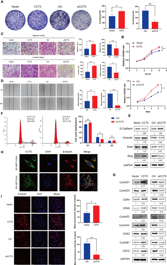
Figure 8.
Role of CCT5 in the proliferation, migration, invasion, and cell cycle regulation of HCC cells. (A) Colony formation assays showing the role of CCT5 on SK-HEP1 cell proliferation. (B) Cell viability of SK-HEP1cell line was determined by CCK-8 assays. (C) The effects of CCT5 on cell migration and invasion were determined by transwell assays in SK-HEP1 cell line. (D) The effects of CCT5 on cell migration were determined by wound healing assay in SK-HEP1 cell line. (E) The expression of EMT markers were examined by western blotting with CCT5 overexpression or downregulation. (F) The cell cycle distribution of SK-HEP1 cells were performed by flow cytometry analyses. (G) The relative expression levels of cyclin D1, cyclin D3, CKD4, CDK6, cyclin E2, cyclin A2, CDK2, cyclin B1, and CDC2 were examined by Western blotting in SK-HEP1 cells. (H) Fluorescence images show staining of CCT5 (green), DAPI (blue) and β-tubulin(red) in SK-HEP1 cells. Scale bar, 10 µm. (I) Representative images of β-tubulin(red) immunofluorescence staining in CCT5-overexpressing and CCT5-knockdown SK-HEP1 cells. Scale bar, 100 µm. Semi−quantitative analysis of β-tubulin fluorescence intensity using ImageJ software. *p < 0.05, **p < 0.01, ***p < 0.001. ns, no significance.

