Figure 3.
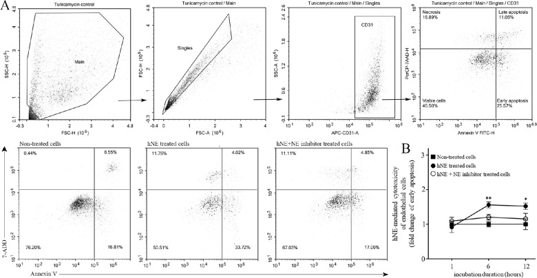
Effect of human NE on mRECs apoptosis. (A) Plots along with gating strategies to determine early and late apoptosis in endothelial cells are depicted. Top panels – tunicamycin (10 µg/mL) was used as positive control to define the quadrants. FSC-H versus SSC-H dot plot (Main) are gated to eliminate debris and then singlet cell (Singles) was selected on FSC-A versus FSC-H. Endothelial cells were gated on CD31 to confirm cell population. Annexin V was used to determine apoptosis and 7-ADD was used to determine cell viability. Bottom panels – Representative flow dot plots from non-treated, hNE treated cells (50 nmol/L) and hNE plus GW311616A inhibitor (GW 150 µMol/L) with quadrants representing endothelial cells in various stages. (B) Early apoptosis is summarized in the graph. All conditions were performed in triplicates. Results are the combination of two experiments. Data are normalized to non-treated cells and expressed as mean ± SD, n = 6 per condition; *P < 0.05, **P < 0.01.

