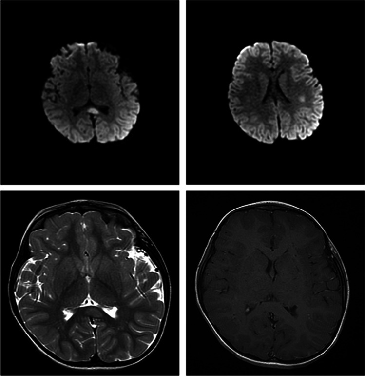
Fig. 3.
Magnetic resonance imaging (MRI) of brain. Axial DWI images show an hyperintense focal lesion in the splenium of corpus callosum and an additional area in the left parietal subcortical site (A and B). Axial T2 image of the same lesion of splenium of corpus callosum that appears subtly hyperintense (C). Axial MRI CE does not evidence any pathological enhancement of the lesion (D)

