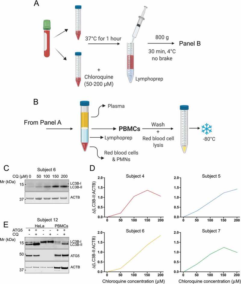
Figure 2.

Autophagic flux in PBMCs can be measured by lysosomal inhibition in whole human blood. (A) CQ was titrated into whole blood samples at final concentrations ranging from 0–200 µM and incubated at 37°C for 1 h before (B) PBMCs were extracted for analysis of LC3B-II by western blot. (C) Western blot for LC3B from PBMCs that were exposed to CQ while in whole blood. (D) Blood from four subjects was processed according to the diagram in panels A and B and is displayed as ΔLC3B-II normalized to ACTB. (E) ATG5 KO HeLa cells that cannot lipidate LC3B-I to form LC3B-II were used to determine that the LC3B antibody used in this study was specific. Mr: molecular weight marker
